Presenters: Mohamed Issa France
This is the case of a 24 y.o male, with no significant past medical or ophthalmological history referred to our centre in Hopital Fondation Ophtalmologique de Rothschild for unsual fundus appearance.
Case Summary:
The patient had a visual acuity of 10/10 both eyes, quiet anterior segment and normal IOP. The fundus exam (cf image) shows a very curious manifestation of diffuse polymorphous concentric fleckoid lesions all over the retina with a perfect macular sparring, good optic nerve head and normal retinal vessels. No areas of atrophy were seen. The main differential diagnosis in front of such fundus exam are : benign familial flecked retina, fundus albipunctatus and retinitis pigmentosa albescens.
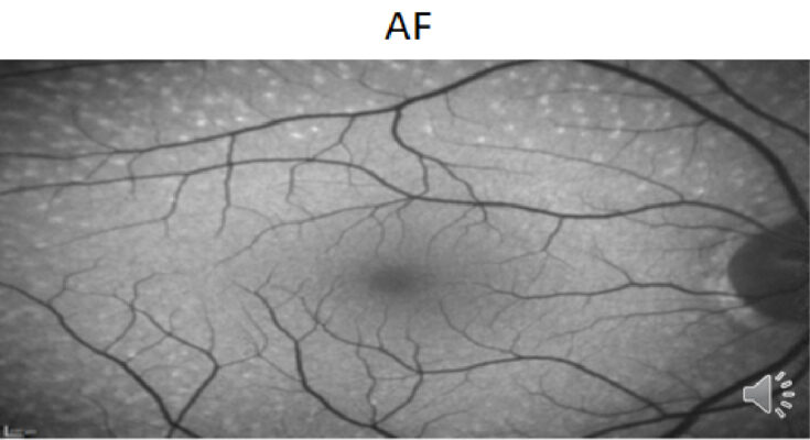
Functionnally speaking, the patient had no particular complaint, no decrease in far or near vision, no nyctalopia, no restriction in visual filed, no dyschromatopsia. No current medication, no history of familial ophthalmological dystrophies. We performed an OCT that locates the lesions at the outer retina and RPE (cf image 2.) The Fundus autofluorescence (cf image 3) shows the moderately hyperautofluorescent behaviour of these « flecks » .
The main exam that helps us distinguish between our differential diagnoses was the global ERG. It came back normal on scotopic and photopic sequences even without prolonged dark adaptation. In front of the absence of clinical symptoms, the normal electrophysiologic exams, we concluded with a diagnosis of Benign familial flecked retina.
However, the genetic results came back, they detected a mutation with a novel variation of the RLBP1 gene, commonly found in the fundus albipunctatus syndrome and retinitis pigmentosa albescens. And this finding made the case even more curious : on one hand, the patient is doing very well on the functional level, on the other hand the genetics detected a mutation with a relatively bad prognosis .
Continuous observation and regular exams are needed in the case of our patient in order to detect as early as possible a clinical or electrophysiological alteration.
Key Images:


Presented during EURETINA Case Club – Series 1, Episode 3 with Prof Ramin Tadayoni (20 Jan, 2022)


